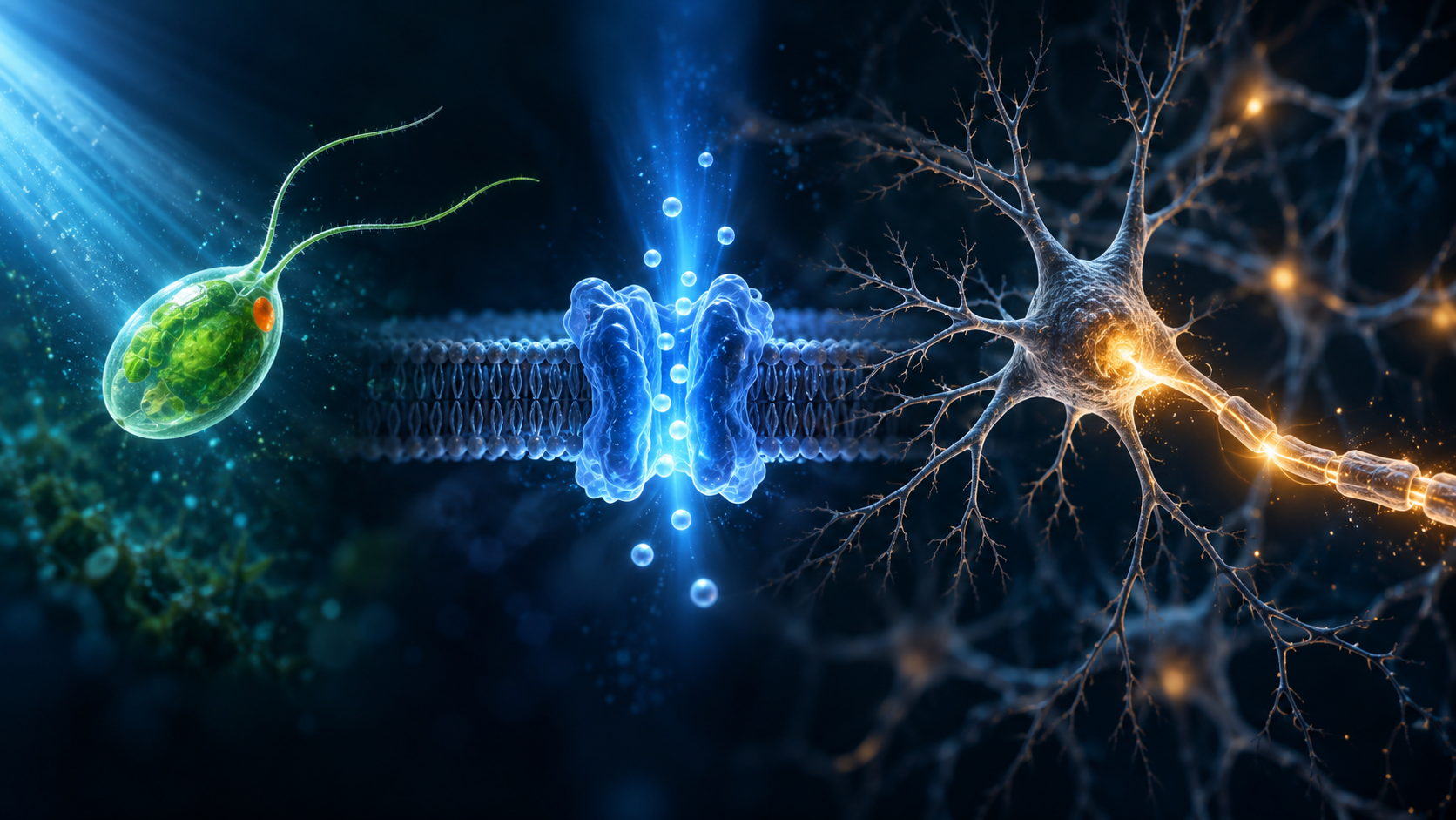
One of neuroscience’s most powerful tools traces back to a cell that spends its life doing something simple: swimming through water and changing course as the light around it changes.
On October 5, 2026, Karl Deisseroth, Peter Hegemann and Georg Nagel were awarded the Nobel Prize in Physiology or Medicine for discoveries involving light-gated ion channels and optogenetics. The prize recognizes a technology that lets researchers use light to manipulate the activity of carefully selected cells, a technique now central to the kind of circuit-level neuroscience behind discoveries about how brain circuits can shape pain and suffering. Its origin, however, wasn’t a brain. It was a single-celled green alga called Chlamydomonas reinhardtii.
Chlamydomonas has two whip-like flagella and an eyespot that helps it respond to light. The organism can alter its swimming direction depending on the light conditions around it, a behavior known as phototaxis. Hegemann spent years trying to understand the molecular machinery behind that response.
That question led researchers to proteins called channelrhodopsins.
In 2002, Nagel, Hegemann and colleagues described channelrhodopsin-1, a light-gated proton channel from Chlamydomonas. A year later, another team led by Nagel characterized channelrhodopsin-2, or ChR2. When blue light struck ChR2, the protein opened a passage through the cell membrane and allowed positively charged ions to cross. Ion channels are the molecular gates that help control electrical signaling in cells, the same broad class of machinery affected by toxins such as those explored in Discvr’s story about an octopus toxin that can stop the heart.
That sounds like a tiny piece of cell biology. It turned out to be the key to something much larger.
The 2003 experiments provided an important clue about how portable the system could be. Researchers put the genetic instructions for ChR2 into frog egg cells and mammalian cells. The protein still worked. Light could open the channel in those foreign cells and alter their electrical state.
No algae needed to be placed inside a brain. Scientists could instead give another cell the instructions for making one of the alga’s light-sensitive proteins.
That distinction is central to understanding optogenetics. The light isn’t magically controlling an ordinary neuron. The target cell first has to express a light-sensitive protein. When the correct wavelength reaches that protein, the channel changes the flow of ions across the membrane. In an excitable cell such as a neuron, that shift can trigger electrical activity.
The leap into neuroscience came quickly.
In 2005, Edward Boyden, Feng Zhang, Ernst Bamberg, Nagel and Deisseroth reported that ChR2 could be expressed in cultured mammalian neurons and used to control their firing with millisecond timing. Neurons communicate through rapid electrical events, so the ability to intervene on a similarly fast timescale gave researchers a new way to investigate what particular cells were actually doing.
How optogenetics turned light into a neuroscience tool
For much of neuroscience, researchers could observe which parts of the brain became active during a behavior and look for patterns. That could reveal an association, but it didn’t always show whether the observed cells were helping cause the behavior.
Optogenetics added a more direct experiment. Researchers could genetically target a defined population of cells, deliver light to them and ask what changed when those cells were activated.
The original ChR2 work was especially useful for exciting neurons. The toolkit soon expanded. In 2007, researchers described a different microbial protein, called NpHR, that could suppress neural firing with light. Combining tools with different optical responses allowed scientists to push activity in opposite directions and probe circuits with far greater precision.
That precision is why optogenetics became so influential. Rather than treating the brain as a collection of broad regions, researchers could begin testing the roles of more specific cell populations and connections. The approach has since been used in animal studies of movement, sleep, fear, memory, addiction and many other functions.
It’s also easy to overstate what the technology does.
Optogenetics isn’t a flashlight that can control an unmodified brain. Cells generally have to be engineered to express the appropriate light-sensitive protein, and researchers still need a way to deliver the right light to those cells. Results in mice or other laboratory animals also don’t automatically translate into treatments for people.
Even the familiar description of optogenetics as an “on-off switch” needs some care. Channelrhodopsin-2 provided a powerful way to activate neurons. Optical inhibition emerged through other microbial proteins and later engineering. The broader field can manipulate activity in both directions, but that capability didn’t come from one protein doing everything.
There is, however, an intriguing human example of where the idea can lead.
In 2021, researchers reported partial recovery of visual function in a blind patient with retinitis pigmentosa. The experimental treatment used a viral vector to make retinal ganglion cells produce ChrimsonR, a light-sensitive protein, along with engineered goggles that converted visual information into light pulses suited to the system.
With the treated eye and the goggles, the patient could perform tasks such as locating, counting and touching objects. Without the goggles, the same visual detection wasn’t demonstrated. The result was partial functional recovery in one patient, not restored normal vision and not proof that optogenetics can broadly cure blindness.
That distinction captures both the promise and the current limits of the field. Optogenetics is already a major research tool. Its medical applications are still developing, and the path from controlling a cell in an experiment to safely treating a human disorder can be long.
The most striking part of the Nobel story may therefore be its starting point.
The researchers who studied Chlamydomonas were trying to understand how a microscopic organism responds to light. The answer revealed a molecular mechanism that could be transferred into other cells. Neuroscientists then turned that mechanism into a way to intervene in neural circuits with extraordinary timing.
A green alga swimming through water didn’t contain a ready-made technology for understanding the brain. It contained a biological solution to a very specific problem. Scientists recognized what that solution could do, carried it into new kinds of cells and kept asking what became possible next.
More than two decades after the first channelrhodopsin papers, that chain of questions has led to a Nobel Prize and to a tool used across neuroscience. The alga is still doing what it evolved to do: responding to light. What changed was our ability to borrow its molecular machinery and use light to ask the brain much sharper questions.



Leave a comment